


Visikol HISTO Visiko 3D组织透明化试剂现货
- 公司名称 世联博研(北京)科技有限公司
- 品牌 其他品牌
- 型号 Visikol HISTO
- 产地
- 厂商性质 代理商
- 更新时间 2021/5/26 10:33:56
- 访问次数 1306
联系方式:李胜亮13261877206 查看联系方式
联系我们时请说明是化工仪器网上看到的信息,谢谢!
| 应用领域 | 医疗卫生,生物产业 |
|---|
Visiko 3D组织透明化试剂现货,Visikol HISTO-M,Visikol® TOX,Visikol HISTO-M,3D透明化成像
visikol透明化试剂全系列现货
,包含多种组织透明试剂,易于使用,无需其他辅助装备,适用于骨骼可视化,软组织透明化,3D细胞培育及植物透明化,价格合理,种类多样.

由于生物组织固有的三维特性,使得生命科学的研究与探索,如对脑部神经投射、血管分布以及肿瘤微环境等研究,需基于生物组织三维空间信息而进行深入研究
我们开发了一套产品,为研究人员提供了从未有的组织视图,以增强其特性。使得研究人员能够在3D中对组织进行标记和成像,以获得完整的特征。我们认为我们的产品是革命性的,因为它们允许研究人员将生物成像的范式从传统的2D组织学方法转变为3D方法。在下面,您可以看到我们的产品类别,包括用于全动物组织的组织透明化试剂和试剂盒(Visikol HITO)、3D细胞培养模型,如器官样、球状体和微问题(Visikol HITO-M)、发育和生殖毒理学研究(Visikol TOX)和植物生物学(Visikol for Plant Biology)。
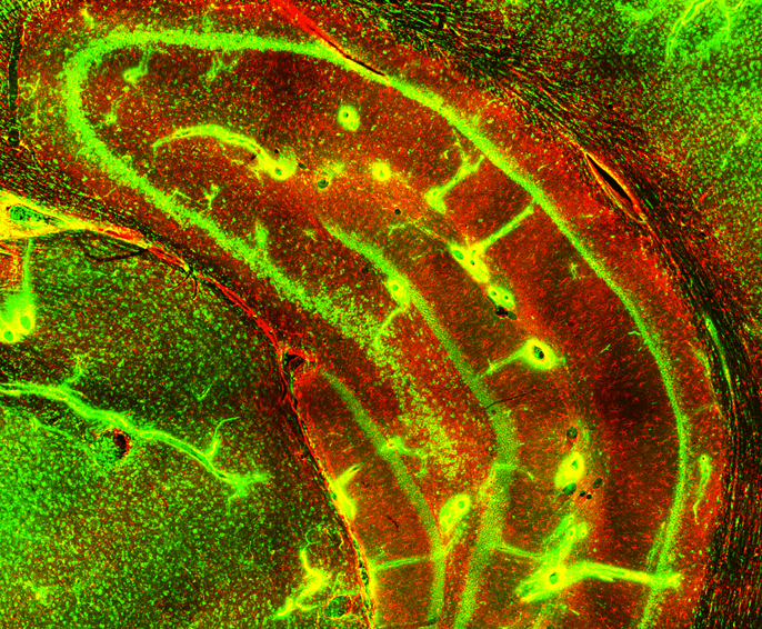
Visikol HISTO
一种快速、易用、可负担且可逆的全组织组织透明化技术。使用共聚焦、光板或双光子显微镜,将荧光蛋白和/或免疫荧光标记与全组织(如小鼠脑、肿瘤)成像的组织透明化剂轻松配对。
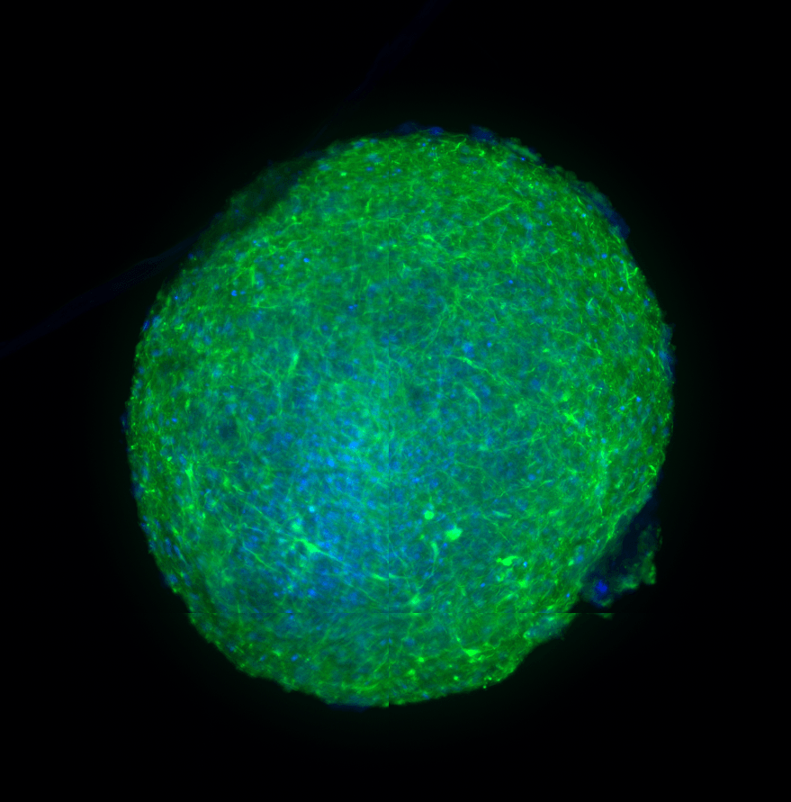
visi kol HITO-M
一种专为高通量自动化分析中的3D细胞培养模型(如器官样细胞、球体、微滴)设计的Visikol HISTO试剂。Visikol HITO-M技术允许完整的3D细胞培养表征,而不是目前仅偏向于外围表征的分析。
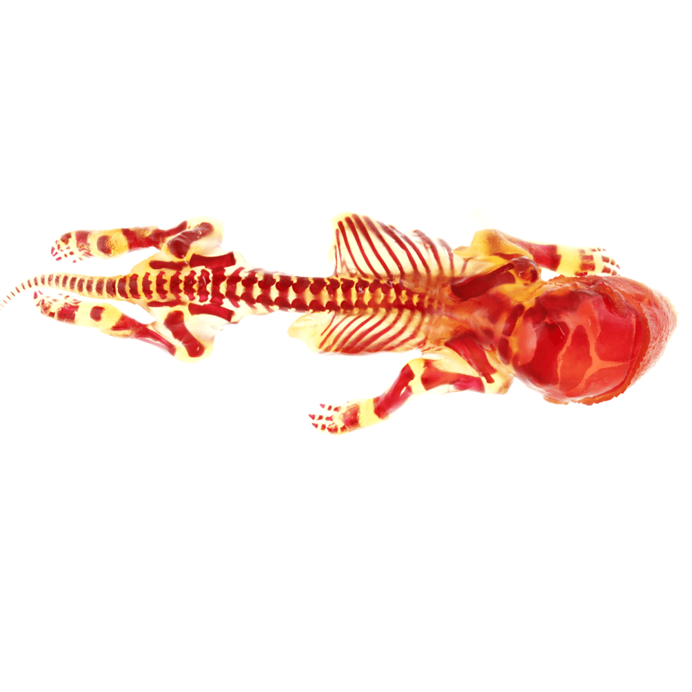
Visikol TOX
一种专门设计用于胎儿骨骼评估的发育和生殖毒理学研究的组织透明化技术。Visikol TOX将这些研究中的骨骼处理时间减少了高达90%,并用非破坏性方法取代了当前基于消化的过程。
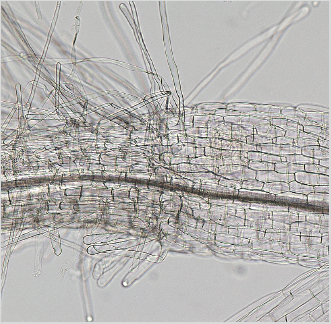
植物生物学巡礼
一种组织透明化技术,设计用于植物样品的显微质量控制。Visikol for Plant Biology通常被用来替代Hertwig的解决方案,该解决方案以前用于此目的,是一种DEA控制的物质。
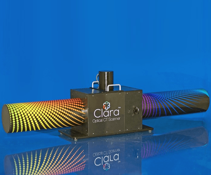
数字灾难援助反应队
我们的数字灾难援助反应队系统将我们的克拉拉光学CT扫描仪与样本自动化、机器学习辅助分析和组织透明化相结合,以3D方式可视化大型组织,如整个鼠标骨骼。光学计算机断层扫描提供了对光板显微镜来说太大的样本成像的能力,并且能够自动评估胎儿骨骼的缺陷,用于灾难援助反应队研究。
Visikol® HISTO™
A rapid, easy-to-use, affordable and reversible tissue clearing technique for whole tissues. Easily pair fluorescent protein and/or immunofluorescent labeling with tissue clearing for whole tissue (e.g. mouse brain, tumor) imaging using confocal, light sheet or 2-photon microscopy.
Visikol® HISTO-M™
A Visikol HISTO reagent designed specifically for use with 3D cell culture models (e.g. organoids, spheroids, microtissues) in high throughput automated assays. The Visikol HISTO-M technology allows for complete 3D cell culture characterization instead of current assays that are biased towards only peripheral characterization.
Visikol® TOX™
A tissue clearing technique designed specifically for use in developmental and reproductive toxicology studies for fetal skeletal evaluation. Visikol TOX reduces skeletal processing time in these studies by up to 90% and replaces the current digestion-based process with a non-destructive approach.
Visikol® for Plant Biology™
A tissue clearing technique designed for use with plant samples for microscopic quality control. Visikol for Plant Biology is commonly used as a replacement for Hertwig’s solution which was previously used for this purpose and is a DEA controlled substance.
Digital DART™
Our Digital DART™ system combines our Clara™ Optical CT scanner with sample automation, machine learning assisted analysis and tissue clearing to visualize large tissues such as a whole mouse skeleton in 3D. Optical CT provides the ability to image samples too large for a light sheet microscope and enables the automated evaluation of fetal skeletons for defects for use in DART studies.


 采购中心
采购中心
 化工仪器网
化工仪器网