 化工仪器网
化工仪器网


详细介绍
带摄像机的primovert hdcam倒置显微镜
达尔文微流体公司与蔡司合作,为您在微流体领域的日常实验设计了Primovert HDCAM装置。
primovert hdcam:一个完整而紧凑的解决方案
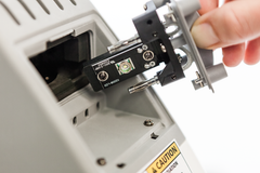
倒置细胞培养显微镜Primovert HDcam设计的终灵活性。primovert能够快速、有效地研究相位对比中的未染色细胞和荧光对比中的荧光细胞。其紧凑的尺寸允许直接在无菌环境下工作,并体验将受污染或敏感材料放入层流柜的安全性。
连接良好的细胞培养实验室
使用LabScope for iPad拍摄图像和视频
利用Primovert HDCam上的众多接口,释放iPad的LabScope Imaging应用程序的功能,将Primovert转换为具有无线成像系统的集成高清相机。LabScope比以往任何时候都更容易捕获显微镜样本的图像和录制视频。创建笔记和报告,编辑图像并将文件保存在Windows网络上。或者同样简单,随时随地与同事分享。由于Primovert HDCAM的接口,您可以通过HDMI将显微镜连接到显示器,并将结果传输和集中显示。从远处可以看到单元格计算。
释放LabScope Imaging应用程序的功能,将Primovert转换为带有无线成像系统的集成高清相机。无论是在实验室还是教室里,LabScope都比以往任何时候都更容易捕捉显微镜样本的图像和录制视频。创建笔记和报告,编辑图像并将文件保存在Windows网络上。或者同样简单,随时随地与同事分享。直观的用户界面可以让您立即工作并小化学习曲线。
Primovert HDCam iPad控制Primovert HDCam LabScope
通过遥控器或计算机监视显微镜
记录你的调查,不要离开无菌的细胞培养环境,也不要用遥控器拍下你的图像。primovert hdcam允许您捕获显微镜图像、录制视频、创建注释和报告以及编辑图像。将文件保存在Windows网络上,或通过无线设备与同事进行“联合”思考。
利用Primovert HDCAM上的众多接口。免费Zun-Lite成像软件提供了灵活的方式将文件传输到您的PC或笔记本电脑。将图像直接传输到层流柜中的监视器。或者把你的数据保存到展台上的SD卡上。
Primovert计算机Zen Primovert HDCam远程Primovert HDCam SD卡
和你的工作流程一样快:打开它,开始评估——一整天,每天
你那一本正经的高清摄像机随时准备好。只需使用方便的台式开关打开和关闭显微镜。由于集成了LED荧光灯,您可以立即开始工作,而无需预热或冷却。空闲时,它会在15分钟后自动关闭,这是另一个节能功能。Primovert易于使用,运行成本低,而且您也很容易,它的内置摄像头可以让您找到舒适的工作姿势,并在一小时又一小时的时间里保持轻松。
Primovert HDCAM手册
PDF手册(见Primovert HDCAM选项)
扩大你的可能性
Primovert有一个通用的相位滑块用于所有目标。您可以使用ph1进行10倍、20倍和40倍放大,并避免在更改放大倍数时必须调整相位。
Pimovert滑块
LED照明具有使用寿命长、色温稳定等优点。使用LED荧光灯,避免灯的升温、降温和调整。使用恒定亮度。
原始LED照明
您可以使用各种安装框架和阶段调整烧瓶和多井板。对于许多培养皿,你也可以扩大阶段。
原始安装架
应用
hela细胞原代gfp细胞原代
hela细胞。放大20倍,相差u2os细胞,gfp标记放大20倍,荧光对比
用带多个物镜的鼻片改变放大倍数4-40倍,相位环。
Primovert有一个4×机头和一系列目标。您可以使用平面消色差和ld平面消色差物镜,其相位环和放大倍数分别为4×和40×。
利用显微镜培训技术助理和学生。
Primovert HDCAM是为联合观察您的结果而设计的。你可以把一个或几个显微镜连接起来。当您在iPad上使用LabScope Imaging应用程序时,您可以捕获和共享图像。
或者,你可以在笔记本电脑、投影仪和SD卡接口的帮助下使用Primovert HDCam,而不需要iPad。
带摄像机的primovert hdcam倒置显微镜
In partnership with ZEISS, Darwin Microfluidics has designed the best Primovert HDcam setup for your everyday experiments in microfluidics.
Primovert HDcam: a Complete and Compact Solution
The inverted cell culture microscope Primovert HDcam is designed for ultimate flexibility. Primovert enables fast, efficient investigations of unstained cells in phase contrast and fluorescent cells in fluorescence contrast. Its compact size allows to work directly in a sterile environment and experience the safety of working with contaminated or sensitive material into your laminar flow cabinet.
The Well-Connected Cell Culture Lab
Capture images and videos with Labscope for iPad
Take advantage of numerous interfaces on Primovert HDcam and unleash the functionality of the Labscope imaging app for iPad to convert your Primovert into an integrated HD camera with a wireless-enabled imaging system. Labscope makes it easier than ever before to capture images and records videos of your microscope samples. Create notes and reports, edit images and save the files on your Windows network. Or just as easily, share them with colleagues – whenever and wherever you want. Thanks to the interfaces of Primovert HDcam, you connect the microscope to this monitor via HDMI and transfer and display your results centrally. Cell evaluations are visualized from the distance.
Unleash the functionality of the Labscope imaging app to convert your Primovert into an integrated HD camera with a wireless-enabled imaging system. Whether in the lab or classroom, Labscope makes it easier than ever before to capture images and records videos of your microscope samples. Create notes and reports, edit images and save the files on your Windows network. Or just as easily, share them with colleagues – whenever and wherever you want. The intuitive user interface gets you to work immediately and minimizes the learning curve.


Monitor your microscope through the remote control or your computer
Document your investigations without leaving the sterile environment of your cell culture and snap your images by remote control, too. Primovert HDcam lets you capture microscope images, record videos, create notes and reports, and edit images. Save the files on your Windows network or do some “joined-up” thinking with colleagues via wireless devices.
Take advantage of numerous interfaces on Primovert HDcam. The free ZEN lite imaging software provides a flexible means of transferring files to your PC or laptop. Transfer images to a monitor directly in the laminar flow cabinet. Or save your data to an SD card on the stand.


As Rapid as Your Work Flow: Switch It On and Start Evaluating – All Day, Every Day
Your Primovert HDcam is always ready to go. Just use the convenient benchtop switch to turn the microscope on and off. Thanks to the integrated LED fluorescence, you start working right away – without warming up or cooling down. When idle, it shuts itself off automatically after 15 minutes – another energy saving feature. Primovert is easy to use, easy on running costs – and easy on you, too, with its integrated camera that lets you find a comfortable working posture and stay relaxed, hour after hour.

PDF Brochure (see Primovert HDcam option)
Expand Your Possibilities
- Primovert has a universal phase slider for all objectives. You can use Ph1 for 10×, 20× and 40× magnification, and avoid having to adjust the phase position when you change the magnification.

- LED illumination gives you the benefit of long life and stable color temperature. Use LED fluorescence to avoid warming up, cooling down and adjustment of the lamp. Work with constant brightness.
- You can use various mounting frames and stage adjustment for flasks and multi-well plates. For many Petri dishes, you can also expand the stage.

Applications

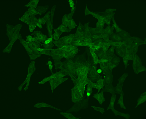
HeLa cells. Magnification 20×, phase contrast - U2OS cells, GFP labeled Magnification 20×, fluorescence contrast
- Use the nosepiece with multiple objectives to change the magnification 4 – 40×, phase ring.
Primovert has a 4× nosepiece and a selection of objectives. You can use Plan-Achromat and LD Plan-Achromat objectives with phase ring and magnifications of 4× and 40×.
- Use the microscope to train technical assistants and students.
Primovert HDcam is designed for the joint observation of your results. You can connect one or several microscopes to each other. When using the Labscope imaging app for iPad, you can capture and share images.
Alternatively, you can use Primovert HDcam without an iPad with the help of laptop, projector and SD card interfaces.
- Capture, edit, document and share results – for example, in quality-management.
Primovert HDcam is designed for the joint observation of your results. You can connect one or several microscopes to each other. When using the Labscope imaging app for iPad, you can capture and share images.
- Use the microscope over several hours.
In automatic mode, Primovert operates in standby. If the device is not used for 15 minutes, it automatically shuts itself off.
Simply press a button to reactivate it.
The ergotube was designed for extended periods of use. You can adjust the viewing height and angle individually to work comfortably in either a seated or standing position.
- Enable several users to operate the microscope.
Primovert HDcam is designed for the joint observation of your results. You can connect one or several microscopes to each other. When using the Labscope imaging app for iPad, you can capture and share images.
- Evaluate unstained, transparent samples such as living cells.
Primovert is equipped with phase contrast. You use a universal phase slider (Ph0, Ph1, and Ph2) for 10×, 20×, and 40× magnification to eliminate the need for adjusting the phase position when adjusting the magnification. Only the Ph1 magnifiers are included within this microscope pack.
- Use the microscope in a sterile environment (laminar flow cabinet in cell culture laboratory).
Primovert’s compact design enables the microscope to fit into any cell culture laboratory. You can put Primovert HDcam straight in your laminar flow cabinet, control it remotely and connect it to a laptop or monitor, thus working directly in a sterile environment.
- Use various cell culture vessels such as petri dishes, multiwell plates and culture flasks.
Primovert comes with a variety of object guides and stages inserts for different cell culture vessels. Use the stage expansion if you want to stack several vessels on the edge. When working with culture flasks, simply remove the condenser.
Primovert is an inverted microscope so it’s easy to observe cells that collect at the bottom of cell culture vessels from below.


 化工仪器网
化工仪器网